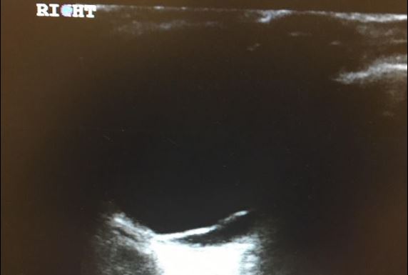
22 year old with no past medical history except for myopia and corrective lenses presents with acute painless monocular vision loss that began 2 days prior. He denies any recent trauma. He’s had a mild intermittent headache since onset, which prompted an outside hospital to order a CT scan, which had no abnormalities and he was discharged. On physical exam his visual fields are intact on the left side, but his right eye has complete deficits in the superior fields. Ultrasound of the right eye yields the following image.
Retinal detachments on ultrasound appear as a hyper-echoic line in the posterior chamber that undulates when the patient looks from side to side.
It’s important to differentiate between “Mac on” and “Mac off” retinal detachments. When the retina has detached off the macula (Mac off), there is little benefit to ophthalmology intervention. When the retina is still attached to the macula (Mac on), urgent intervention is done to preserve vision in that eye and prevent the patient from progressing to a Mac off Detachment. This patient was determined to have a Mac off retinal detachment and thus he was not a candidate for further intervention.
Eric Frendt MS4
References:
– Sonosim Modules: Ocular https://sonosim.ttlms.com/signon/course_launch.cfm?C_ID=4167