Our patient was a 63 year old AA male who was present in the ICU with sepsis requiring pressors. We looked at the standard sites for septic patients: Cardiac, IVC, and Lung. His cardiovascular exam was as we expected: he was known to have a decreased EF and due to his sepsis his IVC was small and collapsed completely on inspiration.
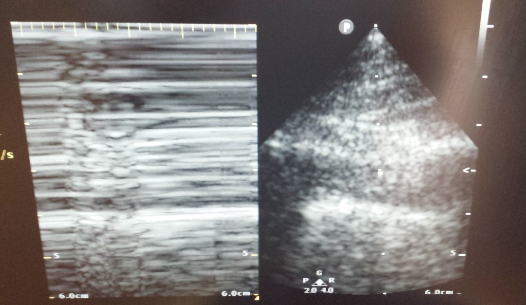
Next we moved on to his lungs. With the linear probe, we began to examine his anterior chest wall where we found bilateral pleural sliding. moving laterally on the right lung, however, we saw something we did not expect: a lack of pleural sliding with no evidence of effusion. How could this be? We saw in the anterior portion of the chest that there was no pneumothorax, yet in a less dependent area we see evidence of pneumothorax. We followed the pleural sliding that was found to lung points inferiorly and laterally and estimated the unaffected portion of the lung to be approximately 8 x 4 cm.
It was later determined that this phenomenon must have been caused by adhesions of the visceral to parietal pleura that prevented the pneumothorax from traveling to the most anterior portion of the chest, creating a loculated pneumothorax.
-Andrew Fellers, MS4
Great case!