6 week old female presenting with recurrence of projectile vomiting. 8 days before current presentation patient had pyloromyotomy after similar clinical presentation. Mother says the surgery went well and patient was doing well at home until yesterday afternoon when she began having projectile vomiting. Patient has vomited about 12 times since then, non-bilious and not bloody. She has not been able to hold down any feedings.
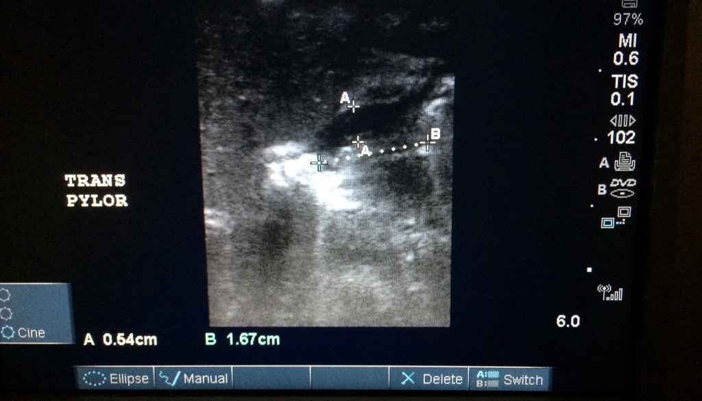
Bedside ultrasound of pyloric sphincter was done. Diameter of single muscular wall on transverse image was 5.4mm (normal <3 mm) and longitudinal measurement of length was 16.7 mm (normal <15 mm). The hypertrophied muscle was found to be hyperechoic.
Ultrasound is the preferred imaging of choice for evaluating pyloric stenosis as it avoids the use of barium, which can be difficult in the pediatric population, it visualizes the pyloric muscle directly, and avoids use of ionizing radiation. Several signs can be noted in pyloric stenosis. Antral nipple sign is seen when pyloric mucosa protrudes into the gastric antrum. Cervix sign is when the pylorus indents into the fluid filled antrum. Target sign is seen when hypertrophied hypoechoic muscle surrounds the echogenic mucosa, seen in the sagittal cross section of the pyloric sphincter.
-Kristina Kyle