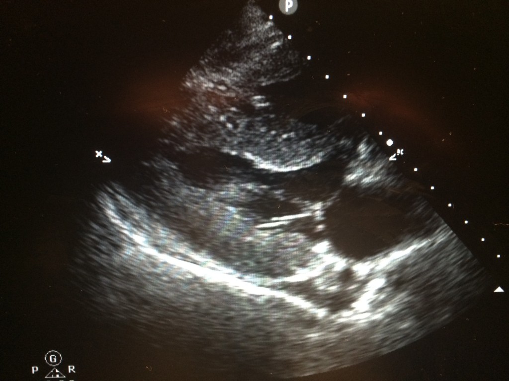
This is a 35 y.o. male I saw while on the MICU service. He originally presented to the Emergency Department with a chief complaint of worsening mental status. Further investigation revealed a history of EtOH cirrhosis and the patient was admitted for hepatic encephalopathy. A parasternal long axis view of the heart in the MICU showed an anechoic area at the posterior left ventricle which was presumed to be fluid accumulation. The fluid is anterior to the descending aorta and posterior to the coronary sinus. No evidence of a swinging heart sign was identified and no diastolic collapse of the right ventricle could be seen. His IVC was greater than 2 cm and demonstrated minimal respirophasic variation. This patient was ventilated at the time this image was taken. Of note, multiple B lines could be seen at R1, R2, L1, and L2. So, does this patient have a pleural effusion or a pericardial effusion?
The key here is structure identification. As can be seen in the image, the fluid is posterior to the coronary sinus but anterior to the descending aorta ( Not well seen in this picture but definitely there). This pattern of fluid accumulation is diagnostic of a pericardial effusion. Had the fluid been posterior to the descending aorta, one could assume that this patient was suffering from a pleural effusion.
Matthew Stevenson, MS4
Excellent post Matt. This is very important especially since in the ICU many of our patients have pleural effusions and differentiating the two is extremely important.
This image shows some hypoechoic structure that appears to me to be posterior to the descending aorta. Wouldn’t this then be considered a pleural effusion, and not a pericardial effusion?
Great observations and questions Danielle! So that hypoechoic structure you are seeing is actually the Coronary Sinus and NOT the descending aorta. This specific patient has a very prominent Coronary sinus.
Visit this page: https://thoracickey.com/the-echocardiographic-examination/
and look at figure 5.10. That shows the difference between descending aorta and coronary sinus (arrow).
There is no IVC on the parasternal axis view so that should not be used as a reference point. Hope that help!!
Or… Should I be using the pericardium as a point of reference and not the inferior vena cava (IVC)?
If I use the IVC as a point of reference, then I would call it a pleural effusion (because the fluid is located posterior to the IVC).
If, on the other hand, I use the pericardium as a point of reference, then I would call it a pericardial effusion (because the fluid is located anterior to the pericardium).
Great observations and questions Danielle! So that hypoechoic structure you are seeing is actually the Coronary Sinus and NOT the descending aorta. This specific patient has a very prominent Coronary sinus.
Visit this page: https://thoracickey.com/the-echocardiographic-examination/
and look at figure 5.10. That shows the difference between descending aorta and coronary sinus (arrow).
There is no IVC on the parasternal axis view so that should not be used as a reference point. Hope that help!!